
Medmont Meridia Corneal Mapping Perth
Corneal mapping is a non-invasive scan that measures the shape and surface of the front of your eye. It can help fit contact lenses, assess astigmatism and monitor keratoconus.



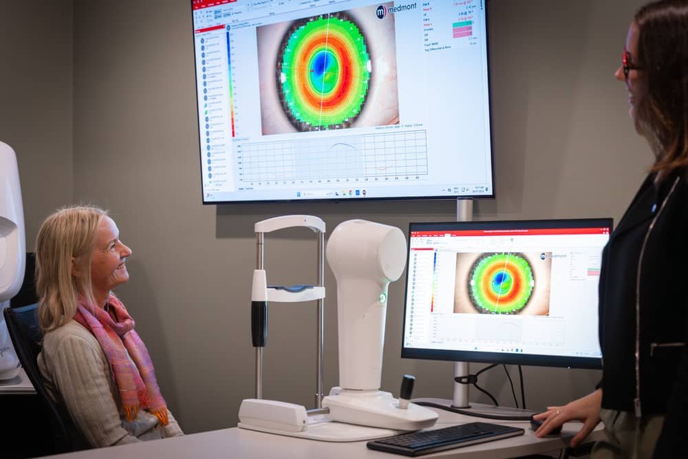
What is corneal mapping?
Corneal mapping is a scan used to measure the shape, curve and surface of the front of your eye.
The front clear window of the eye is called the cornea. Its shape plays an important role in clear vision and comfortable contact lens wear.
At For Eyes, this test is performed using the Medmont Meridia corneal topographer.
This high-resolution imaging system helps our optometrists assess the shape and health of the front of the eye in more detail than a standard exam.
The Medmont Meridia can also support dry eye assessment, tear film review, meibomian gland imaging and contact lens fitting.
When is corneal mapping performed?
Our optometrists may perform corneal mapping during a contact lens fitting, dry eye assessment or diagnostic consultation.
It is especially useful when fitting specialty contact lenses, including toric lenses, rigid gas permeable lenses and orthokeratology lenses.
It may also be used if you have astigmatism, keratoconus, dry eye symptoms or an irregular corneal shape.
Corneal mapping can also help monitor how your eyes respond to contact lens wear over time.
What conditions and applications can this test help with?
The Medmont Meridia helps our optometrists assess the front surface of the eye in detail.
It is especially useful when the exact shape of the cornea matters.
It can assist with:
Contact lens fitting: Helps guide lens type, power and fit based on your eye shape.
Astigmatism assessment: Helps assess irregular corneal shape that may affect vision.
Keratoconus monitoring: Supports assessment of a thinning or cone-shaped cornea.
Orthokeratology fitting: Helps design and monitor overnight lenses that gently reshape the cornea.
Dry eye assessment: Can support tear film and meibomian gland assessment.
Specialty lens fitting: Helps with more complex lens designs, including rigid and scleral lenses.
Monitoring over time: Provides a consistent scan to compare changes between visits.
What should I expect if I have this exam?
Corneal mapping is quick, non-invasive and comfortable.
Nothing touches your eye during the scan.
You will sit at the instrument and look at a target while the machine captures images of the front of your eye.
You may be asked to keep your eye open for a few seconds while the scan is taken.
The test is usually completed quickly and does not usually require eye drops.
Your optometrist will explain the results and how they relate to your vision, contact lenses or dry eye care.
What are the benefits of using the Medmont Meridia in your clinic?
The Medmont Meridia gives our optometrists detailed information about the shape and surface of the eye.
This helps support more accurate contact lens fitting, dry eye assessment and long-term monitoring.
Key benefits include:
High-resolution corneal topography
Detailed assessment of corneal shape and curvature
Useful for specialty contact lens fitting
Supports orthokeratology and rigid lens design
Tear film surface quality analysis
Meibomian gland imaging
Anterior imaging and video
Helps compare changes over time
In simple terms, the Medmont Meridia helps us understand the shape and health of the front of your eye.
This is especially important when fitting contact lenses, assessing dry eye or monitoring corneal conditions.
Need to make an appointment?
Schedule an appointment directly with our clinic or contact us today using our online web form.