
Optos Ultra-Widefield Retinal Imaging Perth
Ultra-widefield retinal imaging is a non-invasive scan that captures a wide picture of the back of your eye. It can help detect and monitor problems like retinal tears, lesions and diabetic eye disease.


What is a ultra-widefield retinal imaging?
Ultra-widefield retinal imaging is a fast, non-invasive scan used to capture a wide image of the back of your eye, called your retina.
At For Eyes, this is performed using Optos ultra-widefield retinal imaging.
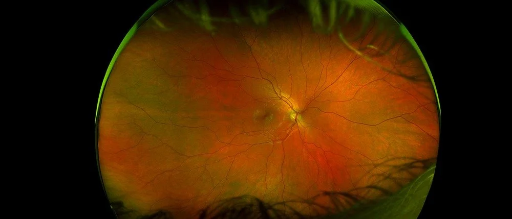
Optos can capture up to 200 degrees, or more than 80%, of the retina in a single image.
This gives our optometrists a much broader view than standard retinal photography.
It is especially useful for seeing the peripheral retina, where some eye conditions can first appear.
When is ultra-widefield retinal imaging performed?
Our optometrists may perform Optos imaging during a routine eye test, diabetic eye examination or diagnostic consultation.
It may be recommended if we need a wider view of the retina, optic nerve, blood vessels or deeper back-of-eye structures.
It is also useful if you have symptoms such as flashes, floaters, sudden vision changes or a shadow in your vision.
Optos imaging may also be used to monitor known retinal conditions or compare changes over time.
What conditions and applications can this test help with?
Optos imaging helps our optometrists assess more of the retina in one image.
This can make it easier to detect changes in the far edges of the retina.
It can assist with:
Retinal tears and detachments: Helps detect peripheral retinal changes that may need urgent care.
Diabetic eye disease: Supports detection and monitoring of diabetic retinopathy and vascular changes.
Vascular occlusions: Helps assess changes in the retinal blood vessels.
Lattice degeneration: Allows wider viewing of thinning or weak areas in the peripheral retina.
Choroidal or retinal lesions: Helps document lesions, naevi or “freckles” at the back of the eye.
Macular and optic nerve assessment: Provides a broad retinal overview alongside other diagnostic testing.
Ongoing monitoring: Sequential imaging allows scans to be compared over time.
What should I expect if I have this exam?
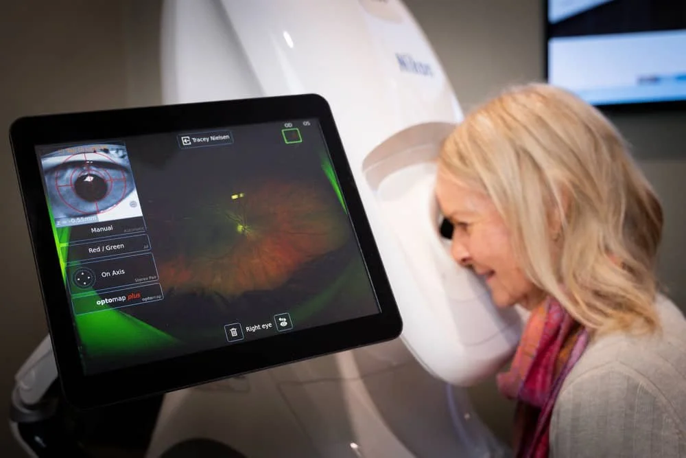
This quick, comfortable scan captures a detailed image of the back of your eye using the Optos Daytona camera. Here's what happens:
You'll be seated at the machine and asked to look into a small opening, focusing on a target light for a few seconds while the scan is taken. Most patients don't need their pupils dilated for this test, so there's no need for drops beforehand and no blurry vision or light sensitivity afterwards — you can drive and go about your day as normal.
The scan itself takes less than a second per eye, and the whole process is usually done within a couple of minutes. It's non-invasive, painless, and involves no contact with your eye.
Once captured, your optometrist will review the image with you on screen, pointing out the optic nerve, blood vessels, and the health of your retina right out to the periphery — areas that aren't always visible with a standard eye check. This image also becomes part of your ongoing eye health record, so any changes can be tracked accurately at future visits.
What are the benefits and clinical usefulness of using Optos ultra-widefield retinal imaging?
Ultra-widefield imaging with the Optos Daytona is valuable because it:
Captures up to 82% of the retina in one image (compared to ~15% with traditional fundus photography), allowing peripheral pathology to be seen that would otherwise be missed
Requires no pupil dilation for most patients, making it faster and more comfortable
Provides a permanent, high-resolution baseline record for monitoring changes over time — useful for diabetic retinopathy, retinal detachment/tears, peripheral degenerations, naevi, and vascular occlusions
Assists early detection of sight-threatening peripheral conditions in routine eye examinations, not just in referred or symptomatic cases
Allows patient education — being able to show patients their own retina in a single wide image is a powerful communication tool
Need to make an appointment?
Schedule an appointment directly with our clinic or contact us today using our online web form.