
Humphrey Visual Field Test Perth
A Humphrey visual field test measures how well you can see across your central and peripheral vision. It helps our optometrists detect and monitor glaucoma, optic nerve conditions and neurological changes affecting the visual pathway.


What is a Humphrey visual field test?
A visual field test measures the complete area you can see while looking straight ahead, including your peripheral or side vision.
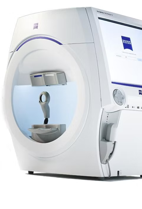
At For Eyes Optometrist, we use the ZEISS Humphrey Field Analyser to perform detailed, automated visual field testing. During the test, small lights appear at different positions and brightness levels. Your responses allow the analyser to create a map showing how sensitive your vision is across the tested area.
Each eye is assessed separately because an eye or neurological condition may affect one eye differently from the other.
Depending on why you are having the test, our optometrist may select a test pattern such as the Central 30-2 Threshold Test. This measures sensitivity at multiple points across the central 30 degrees of your vision.
Visual field testing measures how your vision functions. This makes it an important companion to structural imaging such as Optical Coherence Tomography, which shows the physical condition of the retina and optic nerve.
What does a Humphrey visual field test show?
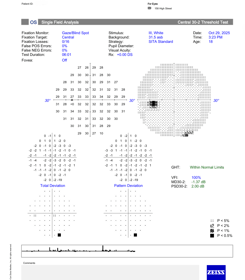
The Humphrey Field Analyser produces a detailed report that helps our optometrists identify areas of reduced visual sensitivity and assess whether the pattern may require further investigation or monitoring.
Your report may include:
Numeric sensitivity values: These show the dimmest light you detected at each tested point. The results are measured in decibels, or dB.
Greyscale map: This presents the same information visually. Darker areas generally represent locations where sensitivity was reduced.
Total Deviation plot: This compares your results with the expected sensitivity for someone of a similar age.
Pattern Deviation plot: This can help identify localised areas of visual field loss after accounting for more general reductions in sensitivity.
Probability symbols: These flag individual test points that fall outside expected limits.
Reliability indices: Fixation losses and false positive or false negative responses help your optometrist determine how dependable the test results are.
Global indices: Measurements such as the Glaucoma Hemifield Test, Visual Field Index, Mean Deviation and Pattern Standard Deviation summarise the overall health and pattern of your visual field.
Your optometrist will interpret these measurements alongside your symptoms, medical history and other eye examination findings. A visual field result is not normally considered in isolation.
When is visual field testing performed?
Our optometrists may recommend visual field testing during a comprehensive eye examination, glaucoma assessment or neurological optometry consultation.
The test may be performed when:
Glaucoma is suspected or has already been diagnosed
Eye pressure or optic nerve findings require further investigation
You have noticed missing, blurred or reduced areas in your peripheral vision
An examination has identified changes involving the optic nerve
Your doctor or specialist has requested an assessment of the visual pathway
You have a neurological condition that may affect vision
Previous visual field results need to be compared over time
Structural imaging and functional testing need to be considered together
Many people do not notice early peripheral vision loss because their central vision may remain clear. Visual field testing can detect changes that may not be obvious during everyday activities.
What conditions and applications can this test help with?
The Humphrey Field Analyser provides an objective map of your visual function. It can assist with the investigation and monitoring of several eye and neurological conditions.
These include:
Glaucoma: Visual field testing can identify patterns of peripheral vision loss associated with glaucoma and help monitor whether the condition is progressing.
Glaucoma suspect and ocular hypertension: Testing may be recommended when eye pressure, optic nerve appearance or other risk factors require closer observation.
Optic nerve conditions: Damage, inflammation or compression affecting the optic nerve may produce recognisable areas of visual field loss.
Neurological conditions: Changes caused by stroke, pituitary lesions or other conditions affecting the brain’s visual pathways may create characteristic field patterns.
Unexplained peripheral vision loss: Testing helps establish where vision has been reduced and whether one or both eyes are affected.
General reductions in visual sensitivity: Cataracts, an uncorrected prescription, a drooping eyelid and other factors can influence test sensitivity and are considered when the results are interpreted.
Ongoing disease monitoring: Repeated tests provide comparable information that can help identify changes over time.
Visual field testing is especially valuable when considered alongside eye pressure measurements, optic nerve examination, OCT scanning and retinal imaging.
What should I expect during a visual field test?
A Humphrey visual field test measures one eye at a time and usually takes around five to six minutes per eye.
You will be seated at the analyser with your chin and forehead supported. One eye will be covered while you look into a bowl-shaped screen and focus on a central target.
Small flashes of light will appear in different areas of your vision. You will hold a response button and press it whenever you notice a flash, even when it appears faint or far to the side.
It is completely normal not to see every light. The test deliberately presents lights that are close to the limits of what you can detect.
There are no air puffs, eye drops or instruments touching your eye. The test should not cause pain or discomfort, although it can feel repetitive.
Try to keep looking at the central target throughout the test. Accurate results depend on steady fixation and natural responses. There is no need to press the button when you are certain no light appeared, but it is fine to respond when you think you may have seen something.
Please let our team know if you need a brief pause or feel uncomfortable at any point.
After the test, your optometrist will review the results with you. We can explain your visual sensitivity map, the reliability of the test and whether the results sit within expected limits for your age.
What are the benefits and clinical uses of Humphrey visual field testing?
Humphrey visual field testing is valuable because it directly measures how you see, rather than only showing how the structures inside your eye look.
Benefits include:
Detecting vision loss you may not have noticed: Early peripheral field changes can develop before they become obvious during everyday activities.
Supporting glaucoma assessment: Recognisable visual field patterns can help with the detection and monitoring of glaucomatous vision loss.
Monitoring changes over time: Repeated tests allow your optometrist to compare results and look for evidence of progression.
Assessing the visual pathway: The pattern of field loss can help identify whether a change may involve the retina, optic nerve or visual pathways through the brain.
Complementing OCT and retinal imaging: Functional results can be compared with structural findings to provide a more complete picture of your eye health.
Checking result reliability: Built-in fixation and response measurements help determine whether the results are dependable or whether the test should be repeated.
Providing standardised records: Each test creates an objective report that can be stored and compared with future examinations.
Helping you understand your results: Your optometrist can show you the visual field map and explain where sensitivity is normal or reduced.
No single test confirms every eye or neurological condition. Your optometrist will consider the visual field results together with your eye examination, symptoms, health history and any other relevant testing.
Need to make an appointment?
Schedule an appointment directly with our clinic or contact us today using our online web form.