GP Co-Management and Complex Vision Diagnostic Support
Our practice has the experienced optometrists and advanced diagnostic equipment to accurately diagnose and manage your patients' complex eye conditions.

How our optometry practice can help your patients
Accurate and appropriate diagnosis of eye health conditions is important. Many ocular emergencies (e.g. red eyes, flashing lights, ocular trauma) require specialised equipment and clinical expertise to accurately diagnose and manage these patients.
Our practice has the advanced equipment and knowledge to ensure patients receive accurate and timely care.
Our optometry services
Paediatric optometry
Myopia management (including axial length measurement)
Complex contact lens fittings (e.g. Orthokeratology and Scleral contact
lenses)
Neurological optometry
Diabetic retinopathy screening
Dry Eye Treatment (including IPL)
Occupational testing: CASA, colour vision, driving standards
Shared care and co-managed outcomes
Currently in Australia, optometrists are able to prescribe topical anti-inflammatory, anti-viral, antibiotic and glaucoma medication. However, some ocular conditions do require oral and/or IV treatment. Our optometrists commonly liaise with GP and/or ophthalmology to facilitate best patient outcomes.
Referral to opthalmology
When a referral to an ophthalmologist is required, waiting times can often be reduced by adding supporting clinical imaging and data from an optometrist.
Acute and complex conditions we can diagnose and treat
-
Red eyes can result from viral, bacterial, allergic, infective, or inflammatory causes — each requiring a different treatment pathway. Accurate diagnosis requires slit lamp assessment and clinical expertise. Our therapeutically endorsed optometrists can identify the cause and initiate appropriate management, including prescribing topical medications where indicated, avoiding unnecessary GP or ED presentations.
-
Foreign bodies, abrasions, and other corneal irritations require prompt, accurate assessment to prevent complications such as infection or scarring. Our slit lamp biomicroscope allows precise evaluation of foreign body depth, rust ring removal, and corneal integrity — ensuring your patients receive timely, appropriate treatment without unnecessary delays or referrals.
-
Painful eyes, flashing lights, floaters, vision reduction, double vision, and ocular headaches can indicate serious underlying pathology requiring urgent assessment. Our advanced diagnostic imaging and clinical expertise allow us to accurately evaluate these presentations, initiate management, and escalate to ophthalmology with supporting clinical data when required.
-
Styes and chalazions are common yet often undertreated eyelid conditions that can cause significant discomfort and recurrence without appropriate management. Our team can accurately diagnose, treat, and advise on ongoing lid hygiene and management strategies, reducing the burden on GP clinics and preventing unnecessary progression or complication.
Accurate and appropriate diagnosis of eye health conditions is important.
Many ocular emergencies (e.g. red eyes, flashing lights, ocular trauma) require specialised equipment and clinical expertise to accurately diagnose and manage these patients.
Our practice has the equipment and knowledge to ensure patients receive accurate and timely care.
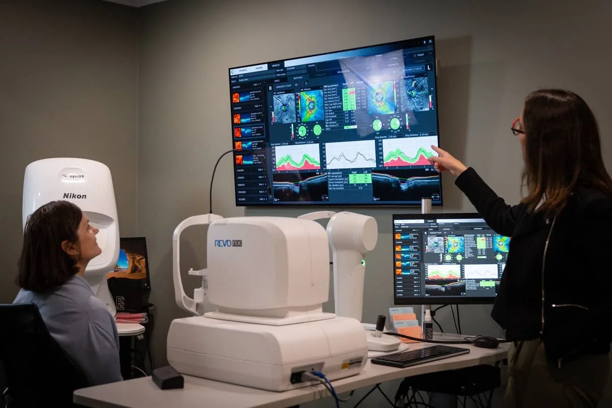
Advanced diagnostics and specialised equipment for complex eye assessments
-
Optical Coherence Tomography (OCT) gives us a cross-sectional view of the retina, macula, optic nerve head and choroidal layers, compared against a normative database.
We use OCT to assess and monitor conditions including glaucoma, macular degeneration, diabetic macular oedema, epiretinal membranes, and optic nerve pathology.
Results are available to include in supporting documentation for ophthalmology referrals.
-
Slit lamp biomicroscopy is essential for accurate diagnosis of anterior segment conditions.
Our slit lamp allows high-magnification, three-dimensional assessment of the cornea, conjunctiva, anterior chamber, iris, and lens.
This is the standard required for diagnosing corneal ulcers, herpetic keratitis, foreign body depth, anterior uveitis, angle closure, and other conditions that cannot be reliably assessed without this equipment.
-
Diabetic eye disease is a leading cause of preventable vision loss, and early detection is critical to preserving sight.
Our diabetic eye examinations include dilated fundus assessment, ultra-widefield retinal imaging and OCT to detect and grade diabetic retinopathy, macular oedema, and other vascular changes.
Findings are documented with clinical imaging and a written report is provided to the referring GP, supporting ongoing shared care and any escalation to ophthalmology where indicated.
-
Our Optos ultra-widefield retinal imaging captures up to 200° of the retina in a single scan — significantly broader than conventional fundus photography — allowing us to detect peripheral pathology that may otherwise go unnoticed.
This includes peripheral retinal tears and detachment, lattice degeneration, peripheral diabetic changes, choroidal lesions and vascular abnormalities.
Imaging is captured rapidly without the need for dilation in most cases, and the results form part of the clinical documentation we provide to referring GPs and ophthalmologists.
Sequential imaging also allows us to monitor and compare changes over time with precision.
At For Eyes Optometrist we have state of the art equipment and a passion for providing excellent eye care. Our optometrists have a wide scope of practice, and all are therapeutically endorsed.
Highly advanced and comprehensive eye examinations
Front of the eye diagnosis
To diagnose most anterior eye conditions, assessment with a slit lamp biomicroscope is required, providing a high-magnification, three- dimensional view of the eye. Some ocular signs that are difficult to diagnose without this technology include:
Foreign body depth & rust ring removal
Corneal infections & ulcers
Viral infections of the cornea (i.e. herpectic)
Anterior angle closure & eye pressure measurements
Iris changes or transillumination
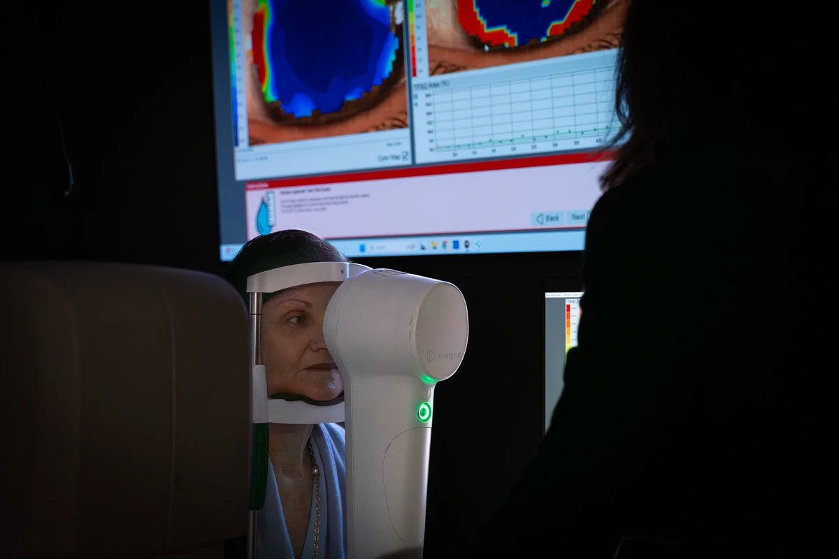
Tear film instability & dry eye
Cataracts & other lenticular changes
We can diagnose all of the above using our in-clinic equipment
Back of the eye diagnosis
To visualise the back of the eye (including the retina, macula, optic nerve, vasculature, and deeper choroidal layers), our optometrists use a ultra-widefield Optos imaging (up to 200 degree scans) and optical coherence tomography with comparison to a normative database. Some ocular signs that are difficult to detect without multimodal imaging or a dilated fundus examination that we have access to include:
Retinal tears and detachment
Vascular occlusions
Diabetic retinopathy & macular oedema
Glaucoma
Age-related macular degeneration
Inherited retinal disease
Ocular melanoma
Inflammation of the posterior segment
Floaters & other vitreous disorders
How our billing works
We made a deliberate decision to move away from bulk-billing to ensure our optometrists have the time and freedom to be thorough. We bill based on time and complexity — like a GP private consultation — which means every test we determine is clinically necessary is included, with no additional charges for imaging or specialist assessments.
What this means in practice:
We spend an average of 40–45 minutes with each patient. That time allows us to complete a genuinely comprehensive examination and feel confident we have the full clinical picture before concluding a consult.
We provide no additional fees and extra charges for any additional tests and assessments — we complete as many as clinically required to fully understand your patient’s condition
Our optometrists have the freedom to investigate as thoroughly as required, without time or billing constraints limiting the scope of the examination
Why choose us?
Thorough clinical reporting — following each appointment, we send a written report to the referring GP with our findings, imaging results, and management plan.
Co-management for complex cases — we're experienced in shared care arrangements for patients requiring both GP and optometry input.
Supporting ophthalmology referrals — we provide clinical imaging and documentation to assist an ophthalmologist's assessment.
Therapeutically endorsed optometrists — all our optometrists hold ophthalmic medicines prescribing endorsement and can initiate topical treatment where indicated.
Advanced diagnostic capability — our clinic is equipped with Optos ultra-widefield retinal imaging, OCT, and slit lamp biomicroscopy for thorough assessment of complex presentations.
Professionally recognised and affiliated — our team are members of Optometry Australia (including Board Member at Optometry Australia WA), ACBO, the Cornea and Contact Lens Society of Australia, and the British College of Optometrists.
Meet the Optometrists
-
![]()
Adrian Rossiter
Founder / Optometrist - BSc(Optom) MOptom MBA, FACBO, IACMM, CASA certified optometrist, Advanced Accreditation in Neuro-Optometric Care, Ophthalmic Medicines Prescriber
Adrian has over 35 years of experience as an optometrist in independent practice, as a contact lens adviser to industry and as a family eye care practitioner. His professional interests revolve around improving children’s vision and working with specialty contact lenses for adults and children.
-
![]()
Sinead Denny
Optometrist - BScOptom, Member of the Australasian College of Behavioural Optometrists, Ophthalmic Medicines Prescriber, Board Member Optometry WA, Clinical Skills Supervisor UWA Doctor of Optometry degree.
Sinead is passionate about all areas of Optometry including contact lenses, myopia control and Indigenous Eye Health. Sinead is also involved in running a Diabetic Eye Screening clinic at Fremantle Hospital and the supervision of students in the Doctor of Optometry at the University of Western Australia.
-
![]()
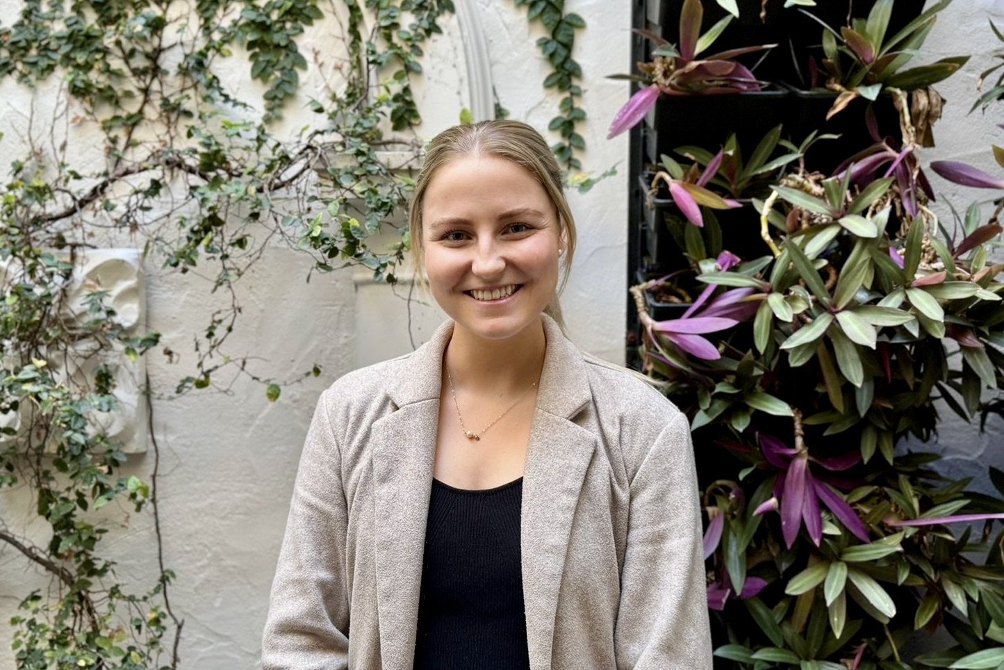
Hannah Jackson
Optometrist - BVisSci, MOptom, AdvCertChVis (ACO), CASA certified optometrist, Opthalmic Medicines Prescriber
Hannah completed her Master of Optometry at Deakin University with distinction. She has a particular interest in paediatric optometry together with colour vision testing and low vision care. She holds an advanced certificate in children’s vision and is a CASA certified optometrist.
-
![]()
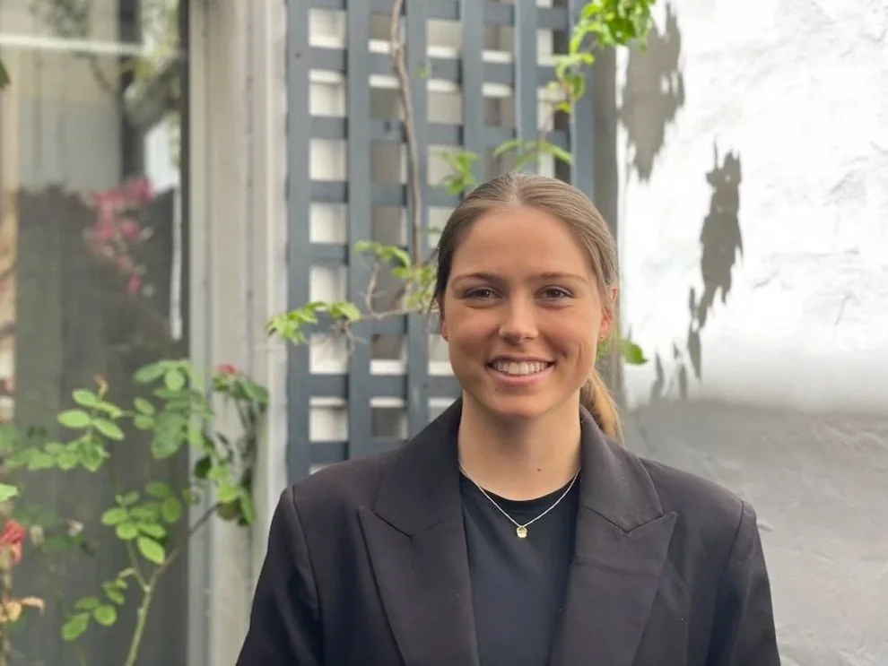
Karega Gibbs
Optometrist - DOptom, BBioMed, Opthalmic Medicines Prescriber
Karega completed her Doctor of Optometry at The University of Western Australia with Distinction. In addition, she has a Bachelor of Biomedical Science (with High Distinction) from the University of Notre Dame. She is passionate about proactive management of eye health including dry eye conditions, and is committed to staying current with advances in optometry, delivering care that is both thorough and compassionate. She is known for a friendly and approachable manner, building trusting relationships with patients and tailoring her care to individual’s needs.