
Optical Coherence Tomography (OCT) Scanning Perth
OCT is a noninvasive imaging method that uses reflected light to create pictures of the back of your eye. It can be used to diagnose and manage diseases like diabetes-related retinopathy and glaucoma.


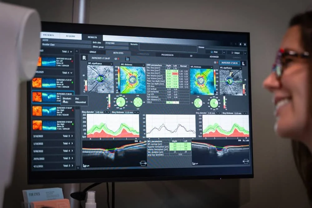
Optical coherence tomography (OCT) is a noninvasive imaging method that uses reflected light to create pictures of the back of your eye. It can be used to diagnose and manage diseases like diabetes-related retinopathy and glaucoma.
Optical Coherence Tomography Explained
Optical coherence tomography, or OCT, is an imaging method used to generate a picture of the back of your eye, called your retina. The noninvasive method produces an image by measuring the amount of dim red light that reflects off of your retina and optic nerve. Optical coherence tomography can measure the thickness of your retina and optic nerve.
Optical Coherence Tomography Explained
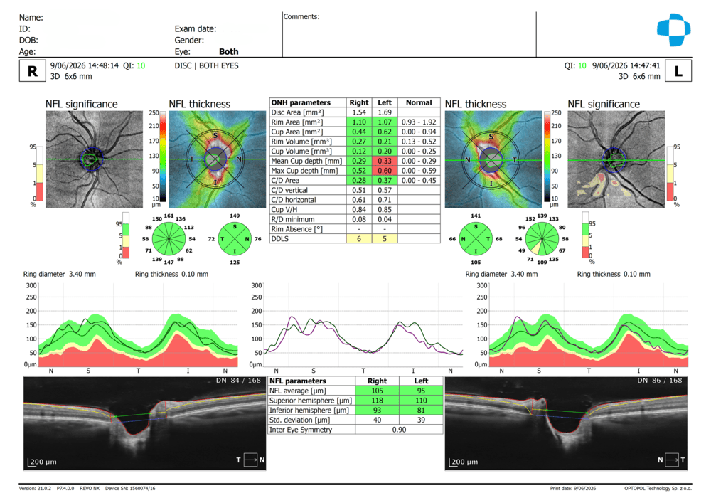
This is an OCT (Optical Coherence Tomography) scan of the Retinal Nerve Fibre Layer (RNFL) and Optic Nerve Head (ONH), captured for both eyes simultaneously using a 3D 6x6mm disc scan protocol. The report includes:
NFL thickness maps – colour-coded (rainbow scale) representations showing nerve fibre layer thickness across the optic disc, with warmer colours (red/orange) indicating thinner areas and cooler colours (green/blue) indicating thicker, healthier tissue
NFL significance maps – comparing the patient's results against a normative database, flagging areas outside the normal range (yellow = borderline, red = abnormal)
ONH parameters – quantitative measurements including disc area, rim area, cup area, cup-to-disc ratios, and DDLS (Disc Damage Likelihood Scale) score
TSNIT curves (Temporal-Superior-Nasal-Inferior-Temporal) – graphs plotting nerve fibre thickness around the optic nerve circumference, compared to age-matched normal ranges
Cross-sectional B-scan images – actual tomographic slices through the optic nerve head showing the physical cup and rim architecture
Clinical Usefulness of OCT
This scan is a cornerstone technology in glaucoma detection and monitoring. Because it provides objective, reproducible, micron-level measurements of the nerve fibre layer, it allows practitioners to:
Detect early, subclinical nerve fibre loss before visual field defects appear
Establish a baseline and track structural change over time (progression analysis)
Differentiate true glaucomatous damage from normal anatomical variation
Correlate structural findings with functional visual field testing for a complete clinical picture
Support treatment decisions (e.g., whether IOP-lowering therapy needs escalating)
When Is Optical Coherence Tomography Performed?
Our optometrists will perform optical coherence tomography in cases where your present with certain conditions at your eye exam or if you already have a condition that they’re helping you manage. Healthcare providers use OCT to diagnose and manage several conditions that affect the eyes, including:
Glaucoma
Age-related macular degeneration
Diabetes-related retinopathy
Cystoid macular edema
Macular pucker
Macular hole
Cone and cone-rod dystrophies
Tumours in your choroid and retina
Monitoring “freckles” (Naevi)
What should I expect if I’m having an optical coherence tomography exam?
This painless scan gives your optometrist a detailed cross-sectional view of your optic nerve and retina, similar to a "CT scan" for the eye. Here's what happens:
You'll be seated at the machine with your chin resting on a chin rest and forehead against a bar to keep your head steady. You'll be asked to look at a fixation target — a small green cross — while the machine takes a series of scans. There are no drops required and no dilation needed for this test, so your vision won't be blurred afterwards and you're free to drive home straight away.
The scan itself only takes a few seconds per eye, using a beam of light (not radiation or X-rays) to build up a detailed image layer by layer. You may need to blink normally between scans, and the machine will usually capture a few images to ensure a clear, high-quality result.
Once complete, your optometrist will review the results with you, showing the thickness and health of your retinal nerve fibre layer compared to normal ranges for your age. This is particularly useful for detecting very early changes in conditions like glaucoma — often before they would be noticeable to you or show up on a standard eye test — and for tracking stability or change over time at future visits.
How often should you have an OCT scan?
Our optometrists will recommend an appropriate review schedule depending on the nature of the condition that is being monitored.
Need to make an appointment?
Schedule an appointment directly with our clinic or contact us today using our online web form.