
How Dry Eye Affects Cataract & LASIK Surgery: What Every Patient Needs to Know
Planning cataract surgery or laser vision correction? Dry eye disease — often undiagnosed — is one of the most important factors affecting your surgical outcome. Here's why it matters, and what needs to happen before you go under the knife.
Key Points at a Glance
Untreated dry eye can cause inaccurate pre-surgical measurements, leading to the wrong intraocular lens (IOL) power in cataract surgery — even when the operation itself goes perfectly.
Both cataract surgery and LASIK can worsen pre-existing dry eye, making ocular surface management before surgery essential to protect your outcome and recovery.
LASIK severs corneal nerves that trigger tear production; patients with moderate-to-severe dry eye may not be suitable LASIK candidates and should be assessed before any surgical planning begins.
The 2025 TFOS DEWS III report identifies dry eye as a critical pre-operative risk factor, not just a post-operative complication.
A simple six-question screening tool (the OSDI-6) can identify patients at risk in under two minutes — it should be part of every pre-surgical eye assessment.
Starting dry eye treatment four to six weeks before surgery allows the ocular surface to stabilise and biometric measurements to be confirmed.
Dry Eye and Eye Surgery: Why the Combination Matters
If you are planning cataract surgery or laser vision correction such as LASIK or SMILE, you have probably thought carefully about the procedure itself — but you may not have considered how the health of your tear film could affect the outcome. Dry eye disease (DED) is one of the most prevalent yet underdiagnosed conditions in eye care, and its interaction with common eye surgeries has significant, well-documented consequences.
In Australia, research suggests that at least one in five adults over 40 has clinically significant dry eye disease — and a large proportion of them do not know it.[4,5] This matters profoundly in a surgical context: an unstable or deficient tear film can distort pre-operative measurements, slow post-operative healing, and in some cases make certain procedures inappropriate altogether.
The key message: Dry eye disease is not just a post-operative complication — it is a pre-operative risk factor. The ocular surface must be assessed and optimised before surgery, not reactively managed afterwards.
The 2025 TFOS DEWS III report — the most comprehensive international update on dry eye disease in eight years, synthesising over 8,000 peer-reviewed studies — specifically identifies ocular surface disease management as a critical component of pre-surgical care for both cataract and refractive surgery patients.[6] Its recommendations, along with growing clinical evidence, now form the basis of best practice for anyone planning eye surgery.
Dry Eye and Cataract Surgery
Cataract surgery is one of the most commonly performed elective procedures in Australia — and one of the most dependent on ocular surface health. The interaction between dry eye and cataract surgery operates at two distinct levels: before the procedure and after it.
Before Cataract Surgery: The Measurement Problem
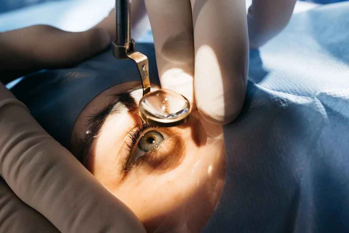
In cataract surgery, the power of the artificial intraocular lens (IOL) to be implanted is calculated using precise measurements of your eye's curvature (keratometry) and length (biometry). The pre-corneal tear film is the first optical surface the measurement instruments encounter — and in dry eye disease, it is unstable and irregular.
A 2025 review presented at the American Society of Cataract and Refractive Surgery (ASCRS) annual meeting outlined the problem directly: a hyperosmolar, unstable tear film skews keratometry and biometry values, leading to inaccurate IOL power calculations.[8] A patient may leave theatre with a technically perfect operation — but end up with residual short-sightedness, long-sightedness, or astigmatism because the measurements taken before surgery were unreliable.
A peer-reviewed study published in 2025 found that treating DED with lipid-containing artificial tears for as little as four weeks before surgery significantly improved the reproducibility of keratometric measurements and reduced refractive prediction error.[9] The clinical takeaway is straightforward: treat the dry eye first, and the measurements become more reliable — and the surgical outcome more predictable.
| Factor | Impact of Untreated DED | Consequence |
|---|---|---|
| Tear film stability | Irregular, fluctuating corneal surface | Inaccurate keratometry readings |
| IOL power calculation | Skewed biometry inputs | Residual refractive error post-surgery |
| Post-operative comfort | Surgery exacerbates pre-existing DED | Significant discomfort, slow recovery |
| Visual quality | Tear film instability causes light scatter | Fluctuating, blurred vision despite successful surgery |
| Patient satisfaction | Unmet vision expectations | Dissatisfaction despite technically good outcome |
After Cataract Surgery: Surgery Can Make Dry Eye Worse
It is equally important to understand that cataract surgery itself is a risk factor for worsening dry eye — even in patients with no prior symptoms. The surgical microscope, operative lighting, corneal incisions (which disrupt the nerves responsible for tear reflex), and the preservatives in post-operative antibiotic and anti-inflammatory drops can all damage the ocular surface and reduce tear production.[10]
In patients with pre-existing DED — even mild or asymptomatic disease — this surgical stress can tip a manageable condition into a significantly symptomatic one. Post-operative dry eye manifests as fluctuating vision, discomfort, grittiness, and prolonged recovery. Identifying and treating DED before surgery creates a buffer: it builds ocular surface resilience and reduces the severity of any post-operative exacerbation.
Dry Eye and LASIK / Refractive Surgery
For patients considering LASIK, SMILE, or PRK for vision correction, the relationship with dry eye is even more direct — and the stakes are higher. Dry eye disease is both a pre-operative screening concern and an established surgical risk factor.
Why LASIK Is a Special Case
LASIK involves creating a thin corneal flap before reshaping the underlying tissue with a laser. This process severs corneal nerves that run close to the surface — nerves that are directly responsible for triggering tear production. Post-LASIK dry eye is one of the most commonly reported complications of the procedure, and it can persist for months or even years in susceptible individuals.
For patients who already have dry eye — even mildly — this nerve disruption can significantly worsen the condition. Symptoms including persistent grittiness, fluctuating vision, light sensitivity, and discomfort can become pronounced after surgery and may affect satisfaction with the outcome even when the refractive correction itself is successful.
Important: Patients with moderate-to-severe dry eye disease may not be suitable LASIK candidates. Your surgeon may recommend an alternative such as PRK (photorefractive keratectomy) or SMILE (small incision lenticule extraction), both of which have a lower impact on corneal nerve density and a more favourable profile for patients with ocular surface disease. This assessment must take place before surgical planning begins.
SMILE and PRK: A Better Option for Dry Eye Patients?
SMILE does not require a corneal flap and disrupts fewer nerves than LASIK, making it associated with lower rates of post-operative dry eye in comparative studies. PRK removes the corneal surface cells (epithelium) rather than creating a flap, which also avoids significant nerve disruption — though recovery is typically slower. For patients with pre-existing DED, these alternatives deserve serious consideration and should be discussed with your surgeon in the context of a thorough ocular surface assessment.
Screening Is Non-Negotiable Before Refractive Surgery
Every patient presenting for a laser vision correction consultation should undergo a comprehensive ocular surface assessment — including tear film analysis, meibography, and validated dry eye screening — before any surgical decision is made. Identifying dry eye at this stage allows it to be treated first, gives the surgeon the information needed to recommend the most appropriate procedure, and ensures that post-operative expectations are realistic and achievable.
What Needs to Happen Before Surgery: A Pre-Operative Roadmap
Guided by the 2025 TFOS DEWS III Management and Therapy Report[6] — the most authoritative international update on dry eye disease, compiled by 80 experts from 18 countries — and current best clinical evidence, the following steps form an effective pre-operative DED management strategy.
Important: Dry eye disease is not just a post-operative complication — it is a pre-operative risk factor. The ocular surface must be assessed and optimised before surgery, not reactively managed afterwards.
Dry Eye Is Treatable — and the Investment Is Worth It
The most encouraging message from the TFOS DEWS III report is that dry eye disease, even when chronic, is manageable. With the right diagnosis and targeted treatment, the ocular surface can be significantly improved. For surgical candidates, this investment in pre-operative care translates directly into better outcomes: more accurate lens calculations, lower rates of post-operative discomfort, more stable vision after surgery, and higher overall patient satisfaction.
At For Eyes Optometrist, we offer comprehensive ocular surface assessment and management as part of our pre-surgical care pathway. If you are planning cataract surgery or laser vision correction, we encourage you to discuss your ocular surface health with us — ideally well before your surgical consultation.
What Needs to Happen Before Surgery: A Pre-Operative Roadmap
Guided by the TFOS DEWS III Management and Therapy Report[6] and current best evidence, the following steps form an effective pre-operative DED management strategy:
Using the OSDI-6 questionnaire as recommended by DEWS III, every patient presenting for cataract or refractive surgery assessment should be screened for dry eye symptoms. A score of ≥4 warrants further investigation and clinical testing prior to biometry.
Non-invasive tear film assessment — including tear break-up time (TBUT), tear meniscus height, lipid layer analysis, and meibography — provides an objective picture of ocular surface health that supplements patient-reported symptoms.
Using the DEWS III driver-based approach, treatment is tailored to the underlying cause. Meibomian gland dysfunction (MGD) — by far the most common driver — is addressed with eyelid hygiene, warm compresses, and in-office treatments such as IPL (intense pulsed light) therapy. Aqueous deficiency is managed with lubricating drops and, where indicated, prescription therapy.
Pre-operative DED management should begin well in advance of any planned surgery — ideally four to six weeks or more — to allow the ocular surface to stabilise and for biometric measurements to be repeated and confirmed.
Co-management between optometrists and the operating surgeon is essential. Your optometrist can provide a formal report on ocular surface status, supporting the surgeon in planning the most appropriate procedure and timing.
Dry Eye Is Treatable — and the Investment Is Worth It
The most encouraging message from the TFOS DEWS III report is that dry eye disease, even when chronic, is manageable. With the right diagnosis and targeted treatment, the ocular surface can be significantly improved. For surgical candidates, this investment in pre-operative care translates directly into better outcomes: more accurate lens calculations, lower rates of post-operative discomfort, more stable vision after surgery, and higher overall patient satisfaction.
At For Eyes Optometrist, we offer comprehensive ocular surface assessment and management as part of our pre-surgical care pathway. If you are planning cataract surgery or laser vision correction, we encourage you to discuss your ocular surface health with us — ideally well before your surgical consultation.
Frequently Asked Questions
Answers to the questions our patients ask us most often about dry eye and eye surgery.
-
Dry eye disease often presents subtly. Common symptoms include a gritty, scratchy, or burning sensation in the eyes, intermittent blurred vision (especially when reading or using screens), excessive watering (a reflex response to dryness), redness, and sensitivity to light or wind.
However, many people with dry eye — including some with clinically significant disease — have no obvious symptoms at all. This is why objective testing by your optometrist is so important, particularly before eye surgery. The OSDI-6 questionnaire used in our clinic takes less than two minutes to complete and can flag those most at risk.
-
Yes — and this is one of the most important messages from the 2025 TFOS DEWS III report. The tear film is the first optical surface of the eye, and an unstable or irregular tear film leads to unreliable pre-operative measurements. The power of your intraocular lens (IOL) is calculated from these measurements, so any inaccuracy can result in residual short-sightedness, long-sightedness, or astigmatism after surgery — even if the operation itself goes perfectly.
In addition, cataract surgery itself can exacerbate dry eye, making pre-operative treatment essential to minimise post-operative discomfort and visual fluctuations.
In addition, cataract surgery itself can exacerbate dry eye, making pre-operative treatment essential to minimise post-operative discomfort and visual fluctuations.
-
Yes. LASIK is one of the most common causes of post-operative dry eye, because the procedure involves creating a corneal flap which severs the corneal nerves responsible for triggering tear production. In patients who already have dry eye — even mildly — LASIK can significantly worsen the condition.
If you have moderate-to-severe DED, LASIK may not be the most appropriate procedure for you, and your surgeon may recommend an alternative such as PRK or SMILE, which have a lower impact on corneal nerves. This is why a thorough ocular surface assessment should be part of every refractive surgery workup.
-
TFOS stands for the Tear Film & Ocular Surface Society, the leading international body dedicated to research and education in tear film and ocular surface science. DEWS (Dry Eye Workshop) is their flagship initiative, producing comprehensive evidence-based reports that set the global standard for understanding and managing dry eye disease.
DEWS III, published in the American Journal of Ophthalmology in June 2025, is the most current and authoritative guidance available. It synthesises more than 8,000 scientific articles published since the previous DEWS II report in 2017, and its recommendations directly shape how we diagnose and treat dry eye disease in clinical practice.
-
Rather than applying the same treatment to every dry eye patient, the driver-based approach involves identifying the specific underlying cause — or causes — of DED in each individual. For example, the most common driver is meibomian gland dysfunction (MGD), in which the oil-producing glands along the eyelid margins become blocked or dysfunctional, leading to rapid tear evaporation. MGD is best treated with eyelid hygiene, warm compression, and in-office treatments like intense pulsed light (IPL) therapy.
Other drivers include aqueous tear deficiency (the eye doesn't produce enough watery component), mucin deficiency, environmental factors, and systemic medications. By targeting the specific driver rather than using a one-size-fits-all approach, treatment outcomes are significantly improved.
-
Current best practice suggests that ocular surface management should begin at least four to six weeks before any planned eye surgery — and ideally longer for moderate-to-severe DED. This allows the tear film to stabilise, biometric measurements to be repeated and confirmed, and any inflammation to settle before the eye is subjected to the additional stress of surgery.
If you are in discussions with a surgeon about cataract or laser vision correction, we recommend scheduling a dry eye assessment with us as early as possible — even before your formal surgical consultation — so that any treatment can begin promptly.
-
We offer a full range of evidence-based dry eye treatments guided by the TFOS DEWS III framework, including:
Eyelid hygiene and warm compression therapy — essential first-line management for meibomian gland dysfunction.
Lubricating eye drops — tailored to your specific tear film deficiency (aqueous, lipid, or mucin-based formulations).
Intense Pulsed Light (IPL) therapy — an in-office treatment for MGD that targets the abnormal blood vessels that drive eyelid inflammation, improving meibomian gland function and reducing evaporative dry eye.
Lifestyle and environmental counselling — addressing screen time, hydration, nutrition (including omega-3 supplementation), and environmental triggers in line with DEWS III recommendations.
Where indicated, we also co-manage with ophthalmologists for prescription-strength anti-inflammatory therapies.
-
Not at all — and this is one of the most important shifts in thinking that TFOS DEWS III reinforces. Dry eye disease is a chronic condition, but it is manageable. With appropriate diagnosis and targeted treatment, most patients achieve meaningful improvement in symptoms, tear film stability, and ocular surface health.
For patients who have previously tried lubricating drops without lasting benefit, it is worth investigating whether an underlying driver — such as meibomian gland dysfunction — is going unaddressed. A thorough ocular surface assessment can reveal the true cause and open the door to more effective treatment.
-
Yes. Dry eye disease — particularly evaporative DED driven by meibomian gland dysfunction — is often not apparent on routine slit-lamp examination, especially early in its course. Dedicated non-invasive testing (tear film break-up time, meibography, tear meniscus measurement, osmolarity) is required to identify subclinical disease that can still meaningfully impact surgical outcomes.
It is also important to note that surgical workup appointments are typically focused on the refractive and anatomical aspects of the eye, and may not include a comprehensive ocular surface assessment. A dedicated appointment with your optometrist fills this gap.
References
Chia EM, Mitchell P, Rochtchina E, et al. Prevalence and associations of dry eye syndrome in an older population: the Blue Mountains Eye Study. Clin Experiment Ophthalmol. 2003;31(3):229–232. PubMed
Chan C. Dry eye disease: current status and improving care. MJA InSight+. 2020. insightplus.mja.com.au
Alcon / Optometry Australia. 2020 Vision Index Report. Cited in: Hospital Health. Dry eye: 77% Australians experience symptoms but many unaware. hospitalhealth.com.au
Australian Eye and Ear Health Survey. Dry eye disease in the Australian Eye and Ear Health Survey. Invest Ophthalmol Vis Sci. 2023. ARVO Journals
Stapleton F, et al. Epidemiology and Risk Factors of Dry Eye Disease: Considerations for Clinical Management. Medicina. 2024;60(9):1458. MDPI
Jones L, Craig JP, Markoulli M, et al. TFOS DEWS III Management and Therapy Report. Am J Ophthalmol. 2025 Jun 2. doi:10.1016/j.ajo.2025.05.039. ajo.com
Stapleton F, quoted in: Healio. ‘The field has advanced tremendously’: TFOS DEWS III offers latest on dry eye disease. December 2025. healio.com
Shah AP. Hyperosmolar tear film and cataract surgery outcomes. Presented at: ASCRS Annual Meeting; April 25–28, 2025; Los Angeles, CA. Reported in: Ophthalmology Times. ophthalmologytimes.com
Bjelobrk M, et al. Impact of Dry Eye Disease and Lipid-Containing Artificial Tears on Keratometric Reproducibility and IOL Calculation in Cataract Patients. NCBI/PMC. 2025. PubMed Central
Sharma N, et al. Understanding and Optimizing Ocular Biometry for Cataract Surgery in Dry Eye Disease: A Review. Semin Ophthalmol. 2023;38(1). tandfonline.com
Planning Eye Surgery? Start Here.
A comprehensive ocular surface assessment before surgery could make all the difference to your outcome. Our team at For Eyes Optometrist is experienced in diagnosing and managing dry eye disease as part of pre-surgical care.
Book an AppointmentFor Eyes Optometrist · Fremantle WA 6160 · Welcoming patients of all ages